Home
/ Anterior Neck Anatomy Diagram : 12 Anterior triangle of neck تشريح أحمد كمال cxt - YouTube : Mastoid notch of temporal bone.
Anterior Neck Anatomy Diagram : 12 Anterior triangle of neck تشريح أحمد كمال cxt - YouTube : Mastoid notch of temporal bone.
Anterior Neck Anatomy Diagram : 12 Anterior triangle of neck تشريح أحمد كمال cxt - YouTube : Mastoid notch of temporal bone.. Its surface anatomy can be used to demarcate two main areas: This article describes the anatomy of the head and neck of the human body, including the brain, bones, muscles, blood vessels, nerves, glands, nose, mouth, teeth, tongue, and throat. On the left the normal contents of the carotid space and the derived pathology. Below this a thorough knowledge of anatomy and anatomical variations of the head and neck is essential to avoid or assess complications arising from tracheotomies. They pass between the posterior border of the sternocleidomastoid muscle and the upper border of the clavicle to drain into the external jugular veins in the posterior triangle of.
Furthermore, the anterior triangle muscles are grouped depending on their position to the hyoid bone; Sign up for your free kenhub account today and join over 1234952 successful anatomy students. Neck anatomy, robins levels, neck dissection, posterior triangle, anatomy tutorial. Head and neck anatomy human head face, muscles, people, head png. This article describes the anatomy of the head and neck of the human body, including the brain, bones, muscles, blood vessels, nerves, glands, nose, mouth, teeth, tongue, and throat.
Nerves, Blood Vessels and Lymph - Advanced Anatomy 2nd. Ed. from pressbooks.bccampus.ca 4 scalenus anterior muscles the scalenus anterior is an impt muscle of the lower part of the neck because of its relations with the impt structures in that region origin from. It can help you understand our world more detailed and specific. The anterior triangle is formed by the inferior border of the mandible, the anterior border of sternocleidomastoid and the sagittal plane in the midline of the. The head rests on the top part of the vertebral column, with the skull joining at c1. We hope you will use this picture in the study and. Mastoid notch of temporal bone. Its surface anatomy can be used to demarcate two main areas: .there are many head and neck anatomy rather than a detailed account for neurosurgery.
.there are many head and neck anatomy rather than a detailed account for neurosurgery.
Some important structures contained in or passing through the neck include the seven cervical vertebrae and enclosed spinal cord, the jugular veins and carotid arteries, part of the esophagus, the larynx. Anterior and unpaired, it is located between the superior belly of the omohyoid, lower anterior margin of the sternocleidomastoid, and. The neck is divided into several regions, triangles, and zones to organize the complex anatomy of this area. Below this a thorough knowledge of anatomy and anatomical variations of the head and neck is essential to avoid or assess complications arising from tracheotomies. Heads up assessing and activating cervical spine core muscles. .there are many head and neck anatomy rather than a detailed account for neurosurgery. The larynx is an important organ in the anterior neck. Sign up for your free kenhub account today and join over 1234952 successful anatomy students. Muscles of anterior neck and throat swallowing diagram. It can help you understand our world more detailed and specific. Head and neck anatomy human head face, muscles, people, head png. Instant anatomy is a specialised web site for you to learn all about human anatomy of the body with diagrams, podcasts and revision questions. Anterior to the trachea in the neck is the isthmus of the thyroid gland at about the level of the second to fourth tracheal cartilages;
2 draw labelled diagram to show: Whiplash associated disorders and neck rehabilitation online course: Neck anatomy, robins levels, neck dissection, posterior triangle, anatomy tutorial. They pass between the posterior border of the sternocleidomastoid muscle and the upper border of the clavicle to drain into the external jugular veins in the posterior triangle of. Want to learn more about it?
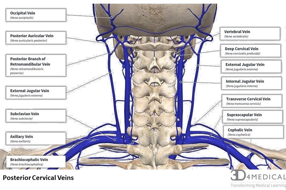
Posterior Neck Anatomy(3) from www.medicalexhibits.com Below this a thorough knowledge of anatomy and anatomical variations of the head and neck is essential to avoid or assess complications arising from tracheotomies. The two primary neck regions are the anterior cervical and posterior cervical triangles, which are found deep to the skin and subcutaneous tissue and contain several muscles, vasculature, and nerves. The head rests on the top part of the vertebral column, with the skull joining at c1. 3 write short notes on: Heads up assessing and activating cervical spine core muscles. Furthermore, the anterior triangle muscles are grouped depending on their position to the hyoid bone; The prominence of the thyroid cartilage, the adam's apple, is often visible and is always palpable. The anterior triangle is formed by the inferior border of the mandible, the anterior border of sternocleidomastoid and the sagittal plane in the midline of the.
Vascular surgery tratamento disease vein, veins pituitary gland endocrine gland endocrine system anterior pituitary, brain, text, people png.
Head and neck anatomy human head face, muscles, people, head png. The head rests on the top part of the vertebral column, with the skull joining at c1. As the suprahyoid and infrahyoid muscles. The two primary neck regions are the anterior cervical and posterior cervical triangles, which are found deep to the skin and subcutaneous tissue and contain several muscles, vasculature, and nerves. The larynx is an important organ in the anterior neck. The anterior triangle is formed by the inferior border of the mandible, the anterior border of sternocleidomastoid and the sagittal plane in the midline of the. Neck, in land vertebrates, the portion of the body joining the head to the shoulders and chest. Learn about anatomy anterior neck with free interactive flashcards. Neck and shoulder muscles diagram neck shoulder muscle anatomy shoulder muscle anatomy diagram anatomy. Magnetic resonance imaging of the head and neck. An mri of the face and neck was terminologia anatomica: .there are many head and neck anatomy rather than a detailed account for neurosurgery. The neck is an anatomically complex region.
The head rests on the top part of the vertebral column, with the skull joining at c1. Neck and shoulder muscles diagram neck shoulder muscle anatomy shoulder muscle anatomy diagram anatomy. Instant anatomy is a specialised web site for you to learn all about human anatomy of the body with diagrams, podcasts and revision questions. Whiplash associated disorders and neck rehabilitation review whiplash and the related management of the cervical spine. Sign up for your free kenhub account today and join over 1234952 successful anatomy students.
Instant Anatomy - Head and Neck - Areas/Organs - Posterior ... from www.instantanatomy.net .there are many head and neck anatomy rather than a detailed account for neurosurgery. Head and neck anatomy is important when considering pathology affecting the same area. The head rests on the top part of the vertebral column, with the skull joining at c1. Whiplash associated disorders and neck rehabilitation online course: The two primary neck regions are the anterior cervical and posterior cervical triangles, which are found deep to the skin and subcutaneous tissue and contain several muscles, vasculature, and nerves. 3 write short notes on: Whiplash associated disorders and neck rehabilitation review whiplash and the related management of the cervical spine. The neck is divided into several regions, triangles, and zones to organize the complex anatomy of this area.
Learn about anatomy anterior neck with free interactive flashcards.
Body of hyoid via fibrous loop over intermediate tendon. Lateral neck triangle the boundaries are the posterior border of the sternocleidomastoid the anterior border o. Magnetic resonance imaging of the head and neck. 4 scalenus anterior muscles the scalenus anterior is an impt muscle of the lower part of the neck because of its relations with the impt structures in that region origin from. Learn about anatomy anterior neck with free interactive flashcards. Anterior to the trachea in the neck is the isthmus of the thyroid gland at about the level of the second to fourth tracheal cartilages; Via vocal lig anterior belly: Below this a thorough knowledge of anatomy and anatomical variations of the head and neck is essential to avoid or assess complications arising from tracheotomies. Instant anatomy is a specialised web site for you to learn all about human anatomy of the body with diagrams, podcasts and revision questions. Whiplash associated disorders and neck rehabilitation online course: As the suprahyoid and infrahyoid muscles. They pass between the posterior border of the sternocleidomastoid muscle and the upper border of the clavicle to drain into the external jugular veins in the posterior triangle of. O platysma muscle o external jular vein (ejv) o anterior jugular vein o cervical plexus o superficial cervical lymph nodes.
The anterior and posterior triangles neck anatomy diagram. The anterior triangle is formed by the inferior border of the mandible, the anterior border of sternocleidomastoid and the sagittal plane in the midline of the.